Skin
Skin Hair & Nails
 Sun Care
 Slimming
 Health
Advantage of Liposomal Technique in Food Supplements
Advantages of the liposomal technique
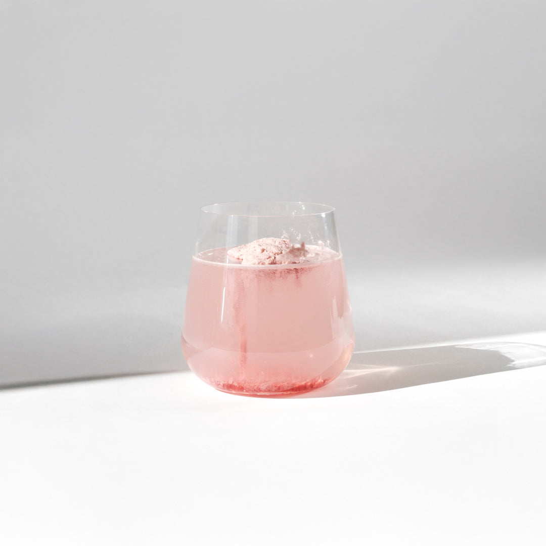
Liposomal technology is based on encapsulating an active ingredient in a liposomal bubble composed of essential fatty acids. This liposomal encapsulation technique is particularly useful for delivering low doses of therapeutic substances in a very targeted manner of the active ingredient, without affecting other parts of the body. The method is often used to benefit from the virtues of Vitamin C, Liposomal Glutathione , or even Curcumin or Vitamin D.
Liposomal: the benefits of microencapsulation
When a capsule is ingested, it must first pass from the mouth through the digestive system and finally be absorbed in the small intestine.
During this process, digestive enzymes in the mouth and stomach, digestive acids, bile salts, and various intestinal flora break down nutrients before they are finally metabolized by the liver and made available to the body. This entire process slows down and reduces the bioavailability of nutrients.
Microencapsulation brings together all the technologies allowing the coating or trapping of active ingredients in solid, liquid or gaseous form within individual particles whose size is between 1µm and 1000µm.
The advantages of this microencapsulation process are numerous, including maximum protection of the active ingredient, particularly against oxidation, light, temperature, or interaction with other components. It is a very useful process for protecting sensitive vitamins such as vitamin C.
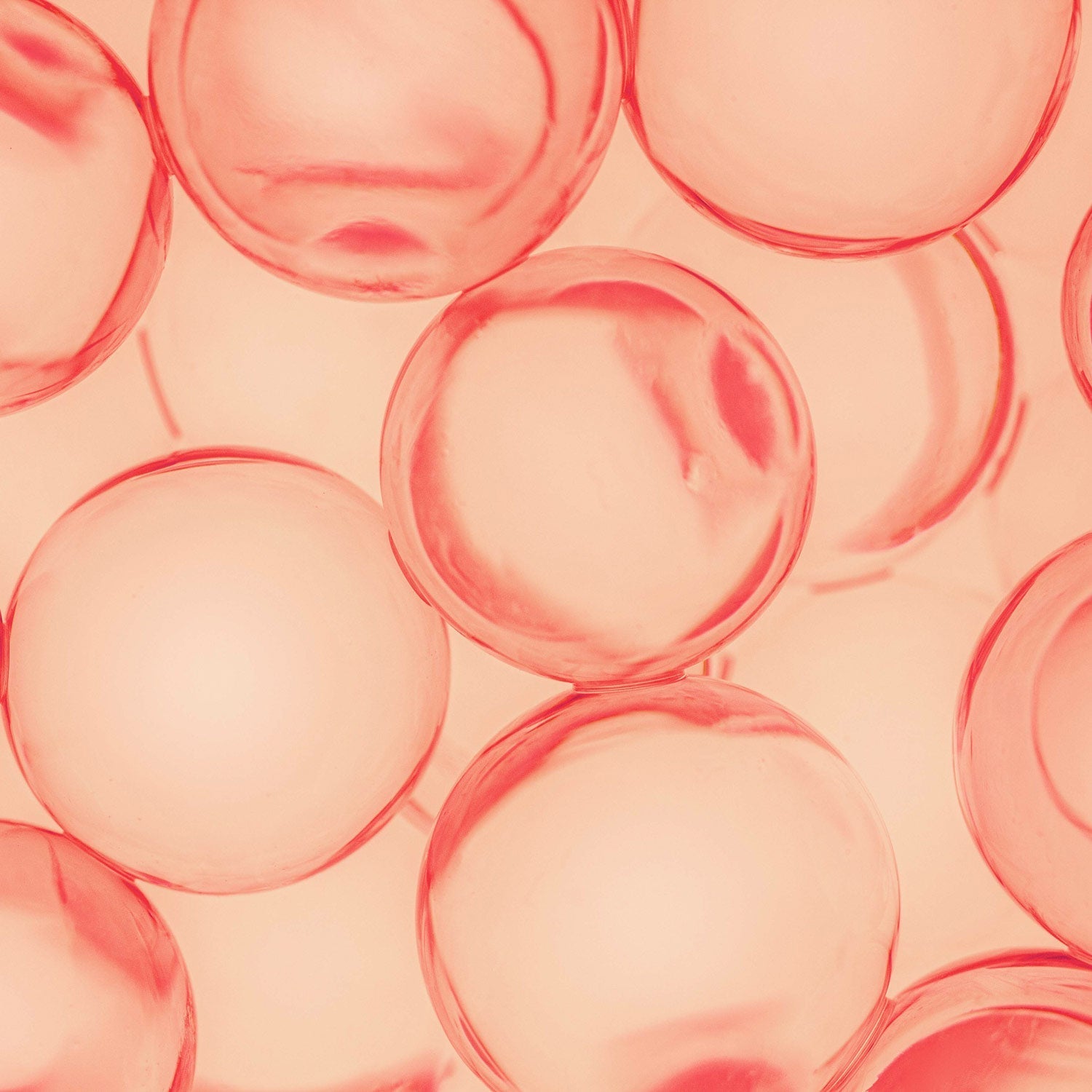
Diagram representing Liposomal technology.
What are liposomes?
Liposomes are encapsulation systems that provide enhanced bioavailability of encapsulated nutrients in the face of environmental, enzymatic, and chemical stresses. These can include the presence of reactive enzymes or chemicals, and exposure to extreme changes in pH, temperature, and ionic strength. This is the technique we use for our Liposomal Vitamin C.
Liposomes are used due to their biocompatibility and biodegradability. Their lack of toxicity, small size, and ability to transport a wide variety of bioactive compounds, thanks to the amphiphilicity of the phospholipid encapsulating material.
Once the liposome reaches the small intestine, it is absorbed by the enterocytes of the villi. Inside the enterocytes, the liposomes are incorporated into chylomicrons.
Once in the bloodstream, liposomes float effortlessly through the lymphatic and circulatory systems. Along the way, they fuse with the body's phospholipid cell membranes, depositing their contents directly into cells; resulting in optimal bioavailability of nutrients such as vitamin D.
Liposome and gastric pH
Gastric pH ranges from 1 to 3, but can reach 4 or 5 in the presence of food. Gastric fluids contain a variety of enzymes such as lipase and protease. The duodenum of the small intestine contains bile salts and many types of enzymes such as amylase, trypsin, and lipase. In addition, the small intestine contains pancreatic enzymes such as pancreatin, trypsin, lipase, peptidase, and maltase. These substances influence the stability of liposomes and encapsulated materials even before they come into contact with intestinal cells.
The mucus layer that lines the surface of the gastrointestinal tract plays an important role in determining the absorption and bioavailability of orally administered drugs. The mucosal surface of the gastrointestinal tract has a very large surface area covered by a monolayer of epithelial cells, connected by tight junctions that present another barrier to drug delivery and can result in rapid elimination of nanoparticles due to rapid cell turnover.
Liposome and microbiota
The mucosal barrier comprises three protective components that provide additional resistance to the gastric mucosal surface. This includes a barrier of compact epithelial cell lining bound by tight junctions, which repel aggressive fluids that can injure the mucosa. The second component is a special mucus blanket secreted by epithelial cells and cells at the mucosal neck. This insoluble mucus forms a protective gel-like coating over the entire surface of the gastric mucosa. The third component consists of bicarbonate ions, also secreted by epithelial cells. When the intestinal microbiota are in symbiosis, the mucosa plays an optimal barrier role.
The different types of liposomes?
In recent years, various attempts have been made to modify the liposomal surface not only to improve their oral stability but also to functionalize the surface of liposomes.
This surface has been modified in several ways by the incorporation of surfactants such as bile salts and Tween 80, or by coating with functional polymers such as chitosan and polyethylene glycol.
Such modifications produce major changes in the various physicochemical properties of liposomes and encapsulated materials. For example, chitosan coating can increase not only the stability of liposomes in various biological fluids, including simulated gastric fluids (SGF) and intestinal fluids, but also the mucoadhesive properties, cellular uptake, and drug solubility.
Micelles
In aqueous solution, molecules having both polar and non-polar regions (= amphiphilic molecules) forming aggregates are called micelles.
The length of the nonpolar tail, the nature and size of the polar head, the acidity of the solution, the temperature, and the presence of added salts are the most important factors determining the type of aggregate obtained. If these parameters are modified, it is possible to change the shape and size of the micelles.
Micelles are widely used by manufacturers for their ability to dissolve and move non-polar substances through an aqueous medium, or to transport substances that are often barely soluble in water.
Micellar aggregates only form when the concentration of the amphiphilic molecule reaches a given concentration called the critical micellar concentration (this is the concentration of surfactant* in a medium above which micelles form spontaneously).
Amphiphilic molecules can form micelles not only in water but also in non-polar organic solvents. In such cases, micellar aggregates are called reverse micelles.
Liposomal & cell membrane formation
The phospholipids in the liposomes that have fused into the cell walls are used by that cell to replace the damaged and/or over-saturated lipids in that cell wall. This renews the cell membrane walls. As a result, blood flows more quickly and easily through the bloodstream, especially through the capillaries, because the blood becomes less viscous (less thick and sticky) and less deformable. The cell membranes are more efficient and more fluid, and the saturated fatty acids and cholesterol that have lodged there are eliminated and metabolized.
Author :
Frédéric LEVY - Doctor of Pharmacy